TB incidence is low in cats and very low in dogs. Because bovine TB is a zoonotic disease (i.e. it can be spread from animal to human), where TB is found in pets, APHA or private vets will inform the Local Health Authority so that any risks to human contacts can be investigated.
If your pet is infected with TB it may show signs of disease which include coughing, wheezing and/or weight loss. Lumps, abscesses or bite wounds which fail to heal, especially those around the head and neck, can also be caused by TB and are most frequently seen in infected cats.
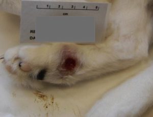
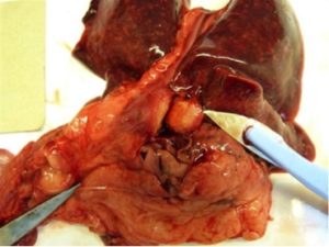
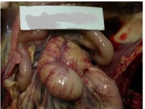
M.bovis infection in cats is predominantly seen as a cutaneous (skin) form. The sites affected are frequently the sites of bite wounds such as the limbs, face and tail head, and the lesions may take the form of raised nodules or non-healing ulcers. The most commonly affected lymph node is the submandibular node. Respiratory and alimentary (gut) forms are seen less frequently. The alimentary form may be seen when cats are fed raw milk from TB infected cows.
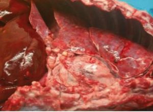
M.bovis infection is much rarer in dogs than cats. It may be associated with bite wounds from infected wildlife. Non-healing bite wounds and enlarged calcified (gritty) lymph nodes have been described. Granulomatous lesions have also been described in the lungs, pleura, liver, kidneys and lymph nodes.
Treatment of TB infected pets is not recommended because of the risk this presents of transmitting the disease to other animals and/or the pet’s owner. If TB is reported in a farm cat or dog, APHA will instigate TB testing of any cattle on the farm and other, potentially exposed cattle, on neighbouring premises.
Testing tissue samples:
There is a legal requirement to report all suspicions of TB infection in the carcases of farmed animals and pets to APHA. Confirmation of disease requires laboratory testing of tissue samples and if there is a strong suspicion of TB, APHA will undertake a pathological examination and bacteriological culture.
If samples are sent to a non-APHA laboratory (at the owner’s expense), the laboratory must notify APHA if M. bovis is identified and the laboratory is encouraged to submit samples to APHA for full identification.
Testing in live animals:
There are currently very few reliable tests that can detect TB in dogs and cats while they are still alive.
- Where possible, biopsy samples, fine needle aspirates (FNAs), or bronchoalveolar lavage (BAL) samples should be submitted for PCR testing and/or bacteriological culture. These tests can be performed by APHA or by an appropriate private laboratory to help confirm the diagnosis.
- Blood samples can be submitted for Interferon-gamma release assay (IGRA) available at The University of Edinburgh (previously offered by BioBest).
- Blood samples can be submitted for DPP VetTB Test available at APHA. This test looks for antibodies, indicating exposure. However, it has not been validated for use in dogs and cats, therefore its diagnostic accuracy in this species is currently unknow.
The interpretation of any test results would need to be considered alongside clinical findings and other case-specific information.
You do not need permission to test live companion animals for TB. They are not covered by the TB Orders legislation and therefore the ‘Request to Undertake Private TB Testing of Non-Bovine Species’ form is not required.