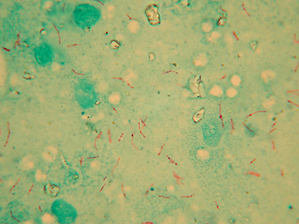
Bovine TB is a chronic, infectious disease caused by the slow-growing bacterium, Mycobacterium bovis (M. bovis). It is mainly a disease of cattle and other bovines but can affect a wide range of species.
Once infected, the opportunity to transmit the disease varies between species. In parts of the UK, there is a recognised reservoir of infection in badgers, with transmission occurring between cattle, between badgers and between the two species. Compared with badgers, wild deer are likely to play a secondary role in the perpetuation of TB in British cattle, and research suggests this occurs only in localised areas at high deer population densities.
Persistence of M. bovis in the environment
Primarily a respiratory disease, transmission of bovine TB normally occurs directly through close contact by breathing in droplets of sputum (thick mucus produced in the lungs) containing M. bovis bacteria exhaled from an infectious animal. Research suggests that M. bovis bacteria can survive in aerosol form for a few hours after exhalation. However, the infection may also be transmitted indirectly, through contact with material (or ingestion of feed) heavily contaminated with M. bovis that may be shed in the sputum, pus, urine, faeces and other excretions of infectious animals. There are practical difficulties with the detection of M. bovis in environmental samples. Nevertheless, the bacterium has been identified in a wide range of sources including soil, faeces, slurry, hay and pasture.
After excretion, M. bovis may remain viable (although it cannot multiply) in the environment for a variable period of time, which can range from a few days to many months depending on the weather and other environmental conditions.
The ultraviolet rays in sunlight have been shown to inactivate M. bovis within hours, although the organism may remain viable for several weeks on pasture. Inactivation is likely to take longer during winter months, as M. bovis survives best in cool, moist environments shaded from direct sunlight. Studies conducted in Ireland have shown that M. bovis can persist in slurry for up to six months, and spreading slurry after storage for less than two months has been associated with an increased risk of bovine TB.
It is not possible to give definitive survival times for M. bovis in different situations, but the following information highlights the underlying factors that enable it to survive in the environment, in order to support the assessment of risk of different circumstances encountered on farm.
Factsheets
Access our factsheets on survival of M. bovis in the environment.
Factors influencing the persistence of M. bovis in the environment
Nature of mycobacteria
M. bovis is a slow growing, very resilient organism notable in its adaptations for survival in the host. It causes an insidious, chronic disease with a variable but often long incubation period with very few symptoms in the early stages of the disease. Depending on the host and other factors, M. bovis is shed by infected animals in sputum, urine, faeces and wound discharges. It is readily transmitted between cattle and susceptible wildlife if they come into close direct contact. However, spread may also occur indirectly if these infected fluids contaminate the environment and then come into contact with susceptible animals in the right conditions. There is evidence from some depopulated farms kept free of cattle for long periods which then suffer recurrence of TB, that M. bovis may persist for prolonged periods in the environment under certain environmental conditions [1]

Adaptation for survival
Pathogenic mycobacteria (those that cause disease) are likely to have arisen from harmless soil dwelling organisms and will have retained many of the adaptations needed for life outside of an animal [2]. M. bovis has further become exquisitely adapted to surviving and flourishing in the hostile environment within macrophages (immune cells). Such adaptations include a waxy cell wall, rich in unique lipid (fat), polysaccharide (carbohydrate) and protein components, and the ability to adopt a quiescent state. This quiescent state, often incorrectly referred to as dormancy, is a state in which bacterial metabolism, replication and its interaction with the exterior is reduced to a minimum.
The structure of the cell wall is fluid and changes in order to adopt these different states. Such adaptations are complex, multi-layered and comprehensive, but result in M. bovis managing to survive within host cells for prolonged periods. These adaptations also confer the ability to survive in the harsh conditions experienced in the environment. Nevertheless, M. bovis does not multiply outside the animal host and it only grows in vitro in selective culture media under special laboratory conditions, and at a very slow rate.
Environmental contamination and survival
Numbers of M. bovis organisms excreted from various sources, in a variety of species, is variable and intermittent. However, as a guide, studies of infected badgers have found 105–106 (106 = one million) organisms/ml in lung exudates and 102–105 organisms/ml in urine and faeces. These numbers are reasonably high, but not as high as may be found with the excretion of other species of bacteria in other diseases. Experimental studies have shown that the infective dose required to establish disease in cattle via the respiratory route may be as low as a single organism [3]. Moreover, there was no significant difference in the likelihood of becoming infected, the time taken to become skin test positive, or the degree of pathology developed between animals receiving a small or larger number of organisms [3].

However, higher doses are believed to be necessary to cause infection via the oral (digestive) route e.g. when cattle ingest grass, fodder or concentrates contaminated with the bacterium, or when calves are fed raw milk from infected cows with TB mastitis. Therefore it would appear that cattle are quite susceptible and at risk of becoming infected after contact with viable M. bovis cells in the environment, but that the portal of entry and amount of viable bacteria present in the contaminated substrate are important factors.
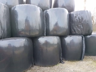
Mycobacteria are very resilient organisms and can survive and remain infective in conditions that would readily kill other bacteria. For example, they are relatively resistant to acid and alkaline conditions and desiccation (drying). However, exposure to UV light and high temperatures from sunlight tend to kill the organisms, whereas cool, moist, dark conditions are protective. Due to the variety of routes of excretion by cattle and wildlife across the farm, contamination of a wide variety of substrates can pose a risk. In general, this includes pasture, fodder and forage, buildings, handling equipment and transport. M. bovis is more likely to be exposed to the drying effects of sunlight on exposed solid surfaces, but survives longer when contamination of softer material such as grass, hay or silage occurs.
Evidence and interpretation of research
There have been a large number of published studies reporting the survival of M. bovis in various substrates and locations [4]. However, it is important to interpret these reports with great care. The experiments are often highly artificial involving, for example, the inoculation of unnaturally large numbers of organisms grown in culture in the laboratory [5]. Such organisms are growing in quite a different biochemical state suited to growth in culture and cannot rapidly adapt to the state required for survival in the environment. In natural circumstances, pathogenic Mycobacteria surviving outside a host are likely to be in a quiescent state, protected by an enhanced cell wall that has changed in structure in readiness for this ecological niche [6].
On the other hand, failure to culture M. bovis in the laboratory is no guarantee that no infectious organisms are present. For example, some reports using culture suggest that pathogenic Mycobacteria may only remain viable for less than three months in soil, even in winter. However, experiments using genomic detection methods have detected M. bovis in soil 21 months after inoculation [7] or are capable of infecting mice after at least one year [8].
Different substrates
The most common substrates that M. bovis may been found in as a result of contamination by infected cattle or wildlife are pasture, concentrates, hay, silage, soil, faeces and water. Conditions in most of these materials are quite favourable for the survival of M. bovis, with a near neutral pH and protection from drying, excessive heat, and UV radiation. Contamination of silage, however, will expose the organism to pH below 4 and very hypoxic (low oxygen) conditions. M. bovis is well adapted to surviving such conditions as it can rapidly respond, in part by adopting its quiescent state. In this state, the bacterium can survive for long periods whilst remaining viable and able to revert to an active infective state within hours [9]. An interesting and potentially significant factor in the successful persistence of mycobacteria in the environment, is their potential to survive and replicate within amoeba and other single celled organisms that live in the soil, in water and in the surface water film on grass.

These organisms often form resistant cysts as protection against drying, extreme pH and other stressors in the environment, and so serve as a further protective layer for mycobacteria within. This potential risk has only recently been recognised and evidence that it is a significant factor in the epidemiology of bovine tuberculosis is yet to emerge. Contamination of hard surfaces such as yards, buildings, handling equipment and transport occurs, but the survival of M. bovis in such situations exposed to sunlight, drying and cleaning/disinfection is likely to be short.
Conclusion
Pathogenic mycobacteria are thought to have evolved from soil-dwelling organisms and they retain the ability to resist the harsh conditions that they may encounter. They are unusually resistant to low pH and drying, although the high temperatures and UV of sunlight reduce survival. Survival may involve the adoption of a quiescent state from which they can readily emerge and become infective. Infection of protozoa might also play a part in survival in the environment, but this needs further investigation. Relatively large numbers of M. bovis excreted by cattle and wildlife can contaminate a range of substrates, and the ability of M. bovis to persist for long periods in an infective state under the right conditions rendering the indirect transmission of bovine TB a substantial risk.
References
1. Rodrıguez-Hernandez et al. Persistence of Mycobacterium bovis under environmental conditions – is it a real biological risk for cattle? Reviews in Medical Microbiology 2016, 27:20–24. doi: 10.1097/MRM.0000000000000059
2. Smith et al. Myths and misconceptions: the origin and evolution of Mycobacterium tuberculosis. Nature Reviews Microbiology. 2009 Jul;7(7):537-44. doi: 10.1038/nrmicro2165. Epub 2009 Jun 1
3. Dean et al. Minimum infective dose of Mycobacterium bovis in cattle. Infection and Immunology. 2005 Oct; 73(10): 6467–6471. https://doi.org/10.1128/IAI.73.10.6467-6471.2005
4. TB hub factsheets
5. Fine, Amanda E. et al. A Study of the Persistence of Mycobacterium bovis in the Environment under Natural Weather Conditions in Michigan, USA. Veterinary Medicine International 2011 (2011): 765430. PMC. Web. 17 Aug. 2018. doi: 10.4061/2011/765430
6. Alderwick, et al. The Mycobacterial Cell Wall – Peptidoglycan and Arabinogalactan. Cold Spring Harb Perspect Med 2015;5:a021113. doi: 10.1101/cshperspect.a021113
7. Young JS, Gormley E, Wellington EMH. Molecular Detection of Mycobacterium bovis and Mycobacterium bovis BCG (Pasteur) in Soil. Applied and Environmental Microbiology. 2005;71(4):1946-1952. doi:10.1128/AEM.71.4.1946-1952.2005.
8. Ghodbane R, Medie F, Lepidi H, Nappez C, Drancourt M. Molecular detection of Mycobacterium tuberculosis sensu stricto in the soil of Niger. Microbiology 160(3):496-501 doi:10.1099/mic.0.073379-0
9. Gengenbacher and Kaufmann, Mycobacterium tuberculosis: success through dormancy. FEMS Microbiology Reviews, Volume 36, Issue 3, 1 May 2012, Pages 514–532, https://doi.org/10.1111/j.1574-6976.2012.00331.x

Useful resources
- DARDNI Review of bovine TB: a review of badger-to-cattle transmission. Allen AR, Skuce RA, McDowell SWJ (2011).
- DARDNI Review of cattle-to-cattle transmission, risk factors and susceptibility. Skuce RA, Allen AR, McDowell SWJ (2011).
- DAERA Slurry literature review. McCallab L, McNair J, Skuce RA (2014). A review of the potential role of cattle slurry in the spread of bovine tuberculosis.
- Veterinary Medicine International Research. Fine AE, Bolin CA, Gardiner JC, Kaneene JB (2011). Study of the persistence of Mycobacterium bovis in the environment under natural weather conditions in Michigan, USA.
- Assessment of the frequency of Mycobacterium bovis shedding in the faeces of naturally and experimentally TB infected cattle. Palmer S, Williams GA, Brady C, Ryan E, Malczewska K, Bull TJ, Hogarth PJ, Sawyer J (2022).